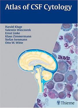

A complete, single-volume reference for the cytological examination of cerebrospinal fluid This full-color atlas presents all the essential information needed for reaching an accurate cytological diagnosis of cerebrospinal fluid and its abnormalities. Designed as a clinical and laboratory reference, "Atlas of CSF Cytology" provides an overview of all the standard diagnostic techniques and offers insight into advanced methods such as flow cytometry and immunocytological phenotyping. Brief descriptions of the indications, advantages, and limitations are provided for each method. An extensive collection of more than 300 high-quality cytological pictures demonstrating normal cell structures, as well as pathological cells in acute and remission phases enables the reader to understand disease processes. Highlights: -Guidelines for the proper handling of specimens, cell preparation, and staining techniques-Review of the common sources of error in diagnosis-Thorough coverage of the techniques for detecting and classifying inflammatory, infectious, neoplastic, and hemorrhagic conditions of the central nervous system-Descriptions of the principle features of cells and the classification of tumor cell types according to current W.H.O. standards-Full-color images depicting pathological alterations of CSF cells -- an indispensable visual aid to comprehension "Atlas of CSF Cytology" is ideal for specialists in neurology, neurosurgery, pathology/neuropathology, cytopathology, microbiology, and laboratory medicine, as well as for those internists, pediatricians, and psychiatrists who frequently request cytological examination of the CSF. Though it is written to meet the needs of specialists, the "Atlas" will also be found accessible and enlightening by interested medical students, interns and residents.
具體描述
讀後感
用戶評價
相關圖書


本站所有內容均為互聯網搜索引擎提供的公開搜索信息,本站不存儲任何數據與內容,任何內容與數據均與本站無關,如有需要請聯繫相關搜索引擎包括但不限於百度,google,bing,sogou 等
© 2025 onlinetoolsland.com All Rights Reserved. 本本书屋 版权所有