Human Brain Anatomy in Computerized Images pdf epub mobi txt 电子书 下载 2026
- CogNeuroSci
- Brain
- Anatomy
- 解剖学
- 大脑
- 医学影像
- 神经科学
- 计算机图像
- 人脑
- 医学
- 神经解剖学
- 影像学
- 医学教育

具体描述
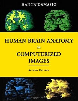
Modern tomographic scans are revealing the structure of the human brain in unprecedented detail. This spectator progress, however, poses a critical problem for neuroscientists and practitioners of brain-related professions: how to find their way in the current tomographic images so as to identify a particular brain site, be it normal or damaged by disease? The problem is made all the more difficult by the large degree of individual neuroanatomical variation. Prepared by a leading expert in advanced brain-imaging techniques, this unique atlas is a guide to the localization of brain structures that illustrates the wide range of neuranatomical variation. It is based on the analysis of 29 normal brain obtained from three-dimensional reconstructions of magnetic resonance scans of living persons. It also provides 177 sections (coronal, axial, and parasagital) of one of those brains so that the same structure presented in the section obtained in one incidence can be identified in the section of another incidence. An additional 209 sections of two incidences of two other brains with different overall configurations are included at the same incidences, so that readers can become familiar with the variability of standard images prompted by different skull shapes. Forty-six normal brains, segmented in to the major lobes, are also included. The atlas is based on a voxel-rendering technique developed in the author's laboratory that permits the reconstruction of the brain in three dimensions. The technique permits the identification of major sulci and gyri with about the same degree of precision that can be achieved at the autopsy table. The volume contains 50 pages of colour illustrations. The second edition of this atlas offers entirely new images, all from new brain specimens. Like the first edition, it will prove to be an essential tool for neurologists, neurosurgeons, neuroradiologists, psychiatrists, and neuroscientists, as well as medical and neuroscience students.
作者简介
目录信息
读后感
评分
评分
评分
评分
用户评价
作为一名对神经科学充满好奇的初学者,我在众多关于大脑的书籍中,对《Human Brain Anatomy in Computerized Images》这本书产生了极大的兴趣。它不像一些枯燥乏味的教科书,而是以一种更加直观、现代的方式呈现大脑的复杂结构。我尤其被书中“Computerized Images”这个关键词所吸引,这暗示着书中会运用大量的医学影像数据,比如CT、MRI等,来揭示大脑的真实解剖细节。我设想,这本书可能会通过将这些二维的影像切片与三维重建的大脑模型相结合,帮助读者建立起一个立体的空间认知。想象一下,能够“亲手”在电脑屏幕上旋转、缩放、剖析一个真实的大脑模型,这本身就是一种令人兴奋的学习体验。而且,我期待书中不仅仅是展示图片,还会配以详尽的文字解释,将每一个脑区、每一条神经纤维的名称、位置、功能及其相互关系一一阐述清楚。或许,它还会穿插一些临床病例的影像,来直观地展示不同疾病对大脑结构的影响,从而加深我们对大脑正常功能的理解。这本书仿佛是一扇窗户,让我们得以窥探人脑这个宇宙中最精密的机器,而我迫不及待地想通过这扇窗户,开始我的探索之旅。
评分作为一名人工智能领域的学生,我对《Human Brain Anatomy in Computerized Images》这本书充满了期待,尤其是在AI与医学影像结合日益紧密的今天。我设想这本书将不仅仅是展示大脑的解剖结构,而是能够将这些结构信息以一种机器可以理解的方式呈现出来。我希望书中能够包含大量的标注数据集,不仅仅是人脑的解剖区域划分,还可能包括神经纤维束的追踪数据,以及不同脑区的体积、形状、纹理等定量信息。这些数据对于训练和优化人工智能模型至关重要,无论是用于疾病诊断、脑功能预测,还是用于开发更智能的神经接口。我期待书中能够探讨如何利用计算机视觉和深度学习技术来自动识别、分割和量化大脑的各个部分,甚至是如何从影像数据中推断出大脑的功能连接和动态变化。如果书中还能提供一些关于如何将这些解剖学知识应用于AI脑模型构建的案例,那将是我学习的巨大财富,能够帮助我更好地理解和构建模拟人脑的AI系统。
评分我是一名热爱科技和前沿技术的新闻记者,我对《Human Brain Anatomy in Computerized Images》这本书的出现感到非常兴奋,因为它代表了科技在揭示生命奥秘方面取得的巨大进步。我预感这本书将是科技与医学交叉领域的一大亮点,它不仅仅是技术展示,更是一种知识的传播和普及。我期待书中能够以通俗易懂的方式,解释计算机成像技术是如何被应用于大脑研究的,比如CT、MRI、PET等技术的原理,以及它们如何“看到”我们肉眼看不见的大脑内部。我希望书中能够展示一些令人惊叹的脑影像图片,这些图片不仅仅是学术研究的成果,更可能成为新闻报道的素材,帮助公众了解大脑的奇妙之处。如果书中还能探讨这项技术在未来医学诊断、治疗甚至人机交互方面的潜力,那将是一篇极具深度和前瞻性的报道主题。这本书,在我看来,是一次科技力量赋能人类自我认知的重要里程碑。
评分我是一名对脑科学充满憧憬的高中生,我一直对人脑充满好奇,但市面上的书籍往往太过专业或晦涩。《Human Brain Anatomy in Computerized Images》这本书,我看到它的名字,便觉得它可能是我理解大脑的最佳入门读物。我设想,书中会用大量的、精美的计算机生成的图像来展示大脑的结构,这些图像可能会像艺术品一样,让我对大脑产生浓厚的兴趣。我希望书中能够用清晰、简洁的语言,介绍大脑的主要区域,比如大脑皮层、小脑、脑干等等,并解释它们各自的作用。如果书中还能穿插一些有趣的小故事,或者介绍一些与大脑相关的科学发现,那会更加吸引我。我期待这本书能够帮助我建立起对大脑的初步认知,激发我对神经科学的进一步学习热情。它不仅仅是一本书,更是我开启脑科学探索之旅的起点。
评分我是一名希望了解大脑衰老和神经退行性疾病的普通读者,我关注《Human Brain Anatomy in Computerized Images》这本书,是因为我希望能够更直观地了解大脑的正常结构,从而更好地理解疾病对大脑的影响。《Human Brain Anatomy in Computerized Images》这本书,在我看来,它提供了一种全新的视角来观察人脑,通过计算机生成的图像,我希望能更清晰地看到大脑的各个部分,以及它们之间的相互联系。我期待书中能够展示健康大脑的影像,并对其结构进行详细的解释。同时,如果书中还能通过对比的方式,展示一些常见神经退行性疾病(如阿尔茨海默病、帕金森病)在计算机图像上的典型变化,并解释这些变化是如何影响大脑功能的,那将对我理解这些疾病非常有帮助。我希望这本书能够以一种易于理解的方式,帮助我认识大脑的脆弱性,并提升我对自己健康的关注。
评分我是一名资深神经影像研究员,阅读了无数篇关于大脑解剖学的文献和专著,而《Human Brain Anatomy in Computerized Images》这本书,恰恰填补了我研究中一直以来渴望的那份“具象化”的空白。在学术研究中,我们经常需要解读大量的MRI、fMRI、DTI等数据,但很多时候,纸面上的解剖图谱和三维模型,即便再精细,也总感觉与那些流动的、动态的影像数据存在一层隔阂。这本书的出现,我预感它将是连接理论解剖与实际影像数据的一个桥梁。我希望书中能够收录最前沿的、高分辨率的脑影像数据,并且不仅仅是静态的图片,而是能够提供交互式的三维模型,允许读者从任何角度、以任何切面来观察大脑的结构。更重要的是,我期待书中能够提供详细的影像学解剖定位方法,以及不同影像序列(如T1、T2、FLAIR、DTI)下特定结构的影像特征对比,这对于我们精确识别病灶、进行脑网络分析至关重要。此外,我希望书中能够包含对常用脑成像技术的简要介绍,以及如何从影像数据中提取和可视化解剖信息的原理,这将极大地提升研究的效率和深度。
评分我是一名对神经解剖学充满热情的艺术史爱好者,我一直对人脑的独特造型和内部结构感到着迷。《Human Brain Anatomy in Computerized Images》这本书,在我看来,它可能不仅仅是一本科学读物,更可能是一本关于“生命之美”的探索。我设想,书中通过计算机成像技术呈现的大脑,将以一种前所未有的清晰度和立体感展现在读者面前。我期待看到那些精妙绝伦的脑沟和脑回的曲线,那些如丝绸般细腻的神经纤维束的交织,甚至是从不同角度观察到的脑血管系统的精巧布局。这本书可能会将科学的严谨性与视觉的冲击力相结合,让读者在理解大脑功能的同时,也能感受到生命体构造的艺术之美。我希望书中能够以一种富有启发性的方式来呈现这些图像,或许会配以文字,解释不同脑区在形态上的独特性,以及这些形态如何与其功能息息相关。这种跨学科的视角,能够让我从艺术的角度去审视大脑,感受生命的鬼斧神工。
评分我是一名临床神经外科医生,每天都在与人脑的复杂结构打交道。《Human Brain Anatomy in Computerized Images》这本书,我看到它的名字,便立刻联想到我日常工作中可能获得的巨大帮助。手术前,我们需要通过CT和MRI来精确规划手术路径,识别肿瘤、血管、重要神经核团的位置,避免损伤关键区域。这本书,如果能将计算机成像技术与人脑解剖学完美结合,我相信它会成为我术前规划的得力助手。我期望书中能够收录大量不同类型、不同视角、不同分辨率的脑影像,并且能够对每一个重要的解剖结构进行清晰的标注,甚至提供不同脑叶、脑沟、脑回的命名和定位标准。我特别希望能看到书中能有针对特定手术区域的详细解剖展示,例如海马体、丘脑、脑干等复杂区域的三维重建和影像特征,这对于我们规避风险、提高手术精度具有至关重要的意义。此外,如果书中还能提供一些常见神经疾病(如脑肿瘤、脑出血、脑梗塞)在计算机图像上的典型表现,并将其与正常解剖结构进行对比,那将极大地提升我们对疾病诊断和治疗的理解。
评分我是一名心理学专业的学生,我一直渴望更深入地理解大脑的结构与心理活动的联系。《Human Brain Anatomy in Computerized Images》这本书,我看到它,就如同看到了一把钥匙,能够帮助我打开通往心理学世界更深层奥秘的大门。我设想,书中不会仅仅罗列大脑的各个部位名称,而是会详细解释每一个脑区在心理活动中扮演的角色,比如额叶在决策和执行功能中的作用,海马体在记忆形成中的重要性,杏仁核在情绪处理上的影响等等。我期待书中能够通过计算机成像技术,直观地展示这些脑区的位置和形态,并且将抽象的心理功能与具体的解剖结构联系起来。如果书中还能包含一些案例研究,比如通过影像学数据来解释某些心理障碍的生理基础,或者展示不同认知任务激活的大脑区域,那将是无价之宝。这本书,我希望它能成为我理解“心智”与“肉体”之间关系的桥梁,让我能够更科学、更深入地探索人类的意识、情感和行为。
评分我是一名退休的生物学教师,对解剖学和生理学有着深厚的兴趣。《Human Brain Anatomy in Computerized Images》这本书,当我得知它利用计算机图像来呈现大脑结构时,我感到非常欣喜。在我的教学生涯中,我一直觉得传统的纸质解剖图谱在立体感和动态性上有所欠缺,而这本书似乎能够弥补这一遗憾。我期待书中能够通过逼真的三维模型,让学习者能够从各个角度清晰地观察大脑的轮廓、内部结构,甚至神经纤维的走向。我希望书中能够提供详尽的图解,解释每一个脑区的功能,以及它们是如何协同工作的。同时,我也期待书中能够运用一些生动的比喻或类比,帮助初学者更容易理解那些复杂的概念。这本书,对我而言,可能是一种重温知识、更新观念的机会,让我能够以一种全新的方式来体验和理解人脑这个神奇的器官。
评分 评分 评分 评分 评分相关图书


本站所有内容均为互联网搜索引擎提供的公开搜索信息,本站不存储任何数据与内容,任何内容与数据均与本站无关,如有需要请联系相关搜索引擎包括但不限于百度,google,bing,sogou 等
© 2026 onlinetoolsland.com All Rights Reserved. 本本书屋 版权所有