

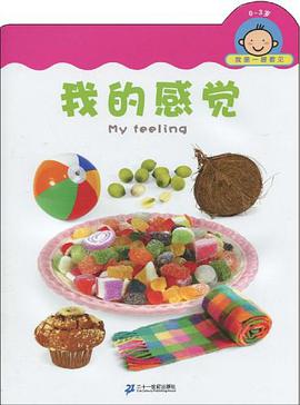
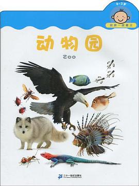
具体描述
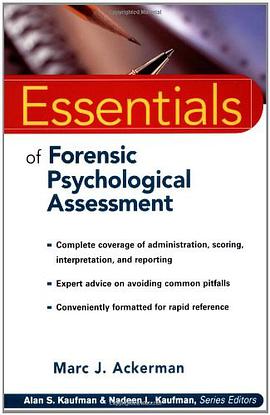
A hundred years ago, a doctor had no way to look within the body of a patient other than to slice it open. That changed radically at the turn of the century, with the discovery of X-rays. X-ray and other forms of diagnostic imaging technology developed slowly but steadily from then until the 1970s, at which point a revolution occurred. Made possible largely by the availability of powerful but inexpensive computers, the rapid and widespread adoption of computed tomography (CT) and, a decade later, of magnetic resonance imaging (MRI) greatly expanded the power of clinical imaging, and even changed the ways in which physicians view and think about the human body. This unique guide explains how the principal imaging devices work and how they help physicians save lives. It gives readers a grasp of the major medical technologies that might come to play important roles in their lives, and it provides succinct, easy-to-understand, and reliable explanations for those who wish to explore the issues of the associated benefits, costs, and risks in an informed manner. In nonspecialized language, Looking Within discusses how X-ray, fluoroscopic, CT, MRI, positron emission tomography (PET), ultrasound, and other medical pictures are created, and explores the essential roles they play in the diagnosis and treatment of patients. It should be of interest to patients and their friends and loved ones, and to those who are simply curious about this vitally important, exciting, and cutting-edge branch of medicine. Its brief but clear descriptions of how these essential tools work should also be of value to health care providers in supporting and educating their patients.
作者简介
目录信息
读后感
评分
评分
评分
评分
用户评价
这本书的叙事方式非常独特,它并非按照传统的逻辑线索来推进,而是更像是一种意识流的呈现,将不同的想法、感悟、观察,巧妙地编织在一起。我发现自己常常会在阅读中,被某个句子所吸引,然后开始进行联想,甚至会突然想起一些被遗忘的童年记忆。这种碎片化的叙事,反而更能引起读者的共鸣,因为人生本身就是由无数个碎片组成的,而这本书就像是将这些碎片,以一种意想不到的方式组合起来,形成一幅完整的画卷。书中的语言充满了诗意和哲理,有些句子读起来,就像是在品味一首优美的诗歌,能够反复咀嚼,每一次都能品出新的味道。我尤其喜欢作者对“当下”的强调,它让我意识到,我们总是活在过去的回忆或者未来的幻想中,而忽略了眼前最真实的生活。这本书就像是一记温柔的提醒,让我重新将注意力拉回到当下,去感受此刻的阳光,去倾听此刻的风声。这种回归当下的能力,让我感觉自己更加充实,也更加快乐。
评分读完这本书,我感到一种前所未有的轻松和释然。它并没有给我灌输什么“成功学”或者“心灵鸡汤”,而是以一种非常自然的方式,帮助我清理内心的杂念,让我的思绪变得更加清晰。我发现自己读这本书时,常常会有“原来是这样”的感悟。那些困扰我许久的某些情绪,在读完某个章节后,突然变得不再那么沉重。作者似乎有一双能够看穿迷雾的眼睛,能够直击问题的本质,并以一种温柔而坚定的方式,引导我们去面对那些不那么美好的部分。我开始更加接纳自己的不完美,也更加理解他人的局限。书中对“放下”的探讨,尤其让我受益匪浅。我总是习惯性地抓住一些过去的事情不放,导致自己无法前进。但这本书让我明白,真正的力量并非在于紧抓不放,而在于懂得适时地放手,然后轻装上阵。这种内心的舒展,让我感觉自己可以更自由地呼吸,也更能以一种平和的心态去面对生活中的起起伏伏。
评分这本书最令我着迷的地方在于其深邃的哲学思考。它并没有用晦涩难懂的语言来包装,而是将一些极具启发性的观点,用一种非常生活化、易于理解的方式呈现出来。我曾几何时,对“意义”这个词感到茫然,觉得它遥不可及,或者只存在于那些伟大的成就之中。但读了这本书,我开始重新审视自己对“意义”的理解。它让我意识到,意义并非宏大的叙事,而是存在于我们日常的点滴之中,存在于每一次真诚的付出,每一次用心的体会。书中的某些章节,让我对时间有了新的认识,不再是单纯的线性流逝,而是充满了无限的可能性和深刻的联系。我开始反思自己过去对时间的浪费,以及如何能更有效地去感知和利用它。作者似乎在不断地抛出问题,引导读者主动去思考,而不是被动接受。这种互动式的阅读体验,让我感觉自己不仅仅是在阅读,更是在参与一场思想的对话。这本书挑战了我固有的认知,也拓宽了我思考的边界,让我对人生有了更宏大的视野和更深刻的理解。
评分这本书的装帧设计非常吸引人,封面采用了深邃的蓝色作为底色,上面点缀着一些细碎的银色星辰,仿佛浩瀚的宇宙在静静地诉说着古老的秘密。书名“Looking within”以一种优雅而内敛的字体呈现,没有过多的修饰,却充满了引人深思的力量。我第一次翻开它,就被那种沉静的氛围所笼罩,仿佛置身于一个远离尘嚣的静谧之地,准备开始一段自我探索的旅程。书页的纸张触感温润,散发着淡淡的墨香,每一页的排版都十分考究,留白恰到好处,让人在阅读时不会感到压迫,反而能更好地沉浸其中。我注意到,书中偶尔会穿插一些手绘的插画,它们风格质朴,线条流畅,为文字增添了几分灵动和意境。这些插画并没有直接解释书中的某个概念,而是以一种更加抽象和象征性的方式,引导读者去体会那些难以言喻的情感和思想。我特别喜欢其中一幅描绘着静谧湖面的画面,湖面映照着远山的剪影,传递出一种宁静而深远的哲学意味。整体而言,这本书在视觉和触觉上都给我带来了非常愉悦的体验,让人迫不及待地想去揭开它文字背后的故事。
评分初读这本书,我最大的感受便是它带来的那种“润物细无声”的力量。它不像有些书那样,上来就抛出很多宏大的理论或者复杂的概念,而是用一种极为温和、细腻的笔触,缓缓地展开。仿佛一位智者,并非直接告诉你答案,而是通过一系列看似寻常的叙述,引导你去发现事物内在的联系和规律。我发现自己常常在阅读的某个瞬间,停下来,陷入沉思,脑海中会涌现出许多与生活息息相关的片段。这些片段并非书中直接提及的内容,而是书中传递出的某种情绪、某种视角,触动了内心深处的回响。书中对人际关系的描绘尤其让我印象深刻,它没有简单地划分对错,而是深入剖析了情感的复杂性和沟通的微妙之处。我能够从中找到很多自己曾经经历过的场景,那些困惑、那些误解,以及最终的和解与成长。作者似乎有一种洞察人心的能力,能够精准地捕捉到那些隐藏在言语之下的真实想法和情感波动。这种细腻的观察和深刻的理解,让我感觉这本书不仅仅是一本书,更像是一位无声的朋友,默默地陪伴着我,分享着我对世界的看法。
评分 评分 评分 评分 评分相关图书


本站所有内容均为互联网搜索引擎提供的公开搜索信息,本站不存储任何数据与内容,任何内容与数据均与本站无关,如有需要请联系相关搜索引擎包括但不限于百度,google,bing,sogou 等
© 2026 onlinetoolsland.com All Rights Reserved. 本本书屋 版权所有